E coli gram stain slide 223110
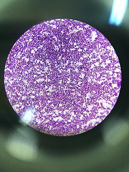
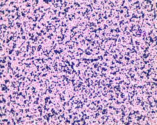
E coli in gram stain E coli in Gram stain showing gram negative rods having size of about µm long and diameter as shown above picture Introduction of Escherichia coli E coli is a Gram negative, aerobe and facultative anaerobic, rodshaped bacteria Optimal temperature for growth is 3617°C with most strains growing over the range 1844 °CA slide is also not acceptable for examination if microorganisms that should be grampositive appear pink This may indicate that either the Gram's iodine was not applied or the slide was overdecolorized Staining grampositive and gramnegative control slides along with the patient's smear would confirm that proper staining technique was usedOn Endo agar it looks like lactose negative)All four strains are mannitol positive (best seen in fig D), cellobiose negative (strains A, B)

Gram Staining Principle Procedure And Results Learn Microbiology Online
E coli gram stain slide
E coli gram stain slide-4 Aseptically transfer Escherichia coli (E coli) to circle on right Allow slide to heat fix S epi D Perform the Gram Stain 1 Take the heat fixed Gram slide to one of the staining racks located at the benches with sinks Flood each slide with crystal violet and allow to stand for one minute Rinse the slide with water 2Both the E coli and S aureus should appear blue/purple What was the purpose of the counterstain?


Http Coltonanderson1 Weebly Com Uploads 2 4 3 0 Manual Pdf
1 Escherichia coli a Fix a smear of Escherichia coli to the slide as follows 1First place a small piece of tape at one end of the slide and label it with the name of the bacterium you will be placing on that slide 2Using the dropper bottle of deionized water found in your staining rack, place 1/2 of a normal sized drop of water on a clean slide by touching the dropper to the slideEscherichia coli (/ ˌ ɛ ʃ ə ˈ r ɪ k i ə ˈ k oʊ l aɪ /), also known as E coli (/ ˌ iː ˈ k oʊ l aɪ /), is a Gramnegative, facultative anaerobic, rodshaped, coliform bacterium of the genus Escherichia that is commonly found in the lower intestine of warmblooded organisms (endotherms) Most E coli strains are harmless, but some serotypes (EPEC, ETEC etc) can cause serious foodAs indicated in the student notes, the first slide that was prepared using aseptic technique and the Gram Staining method was a slide containing cultures of M luteus and E coli When examining this slide under the microscope at 1000X magnification, I observed some clusters and mostly doubles of coccishaped purple colonies
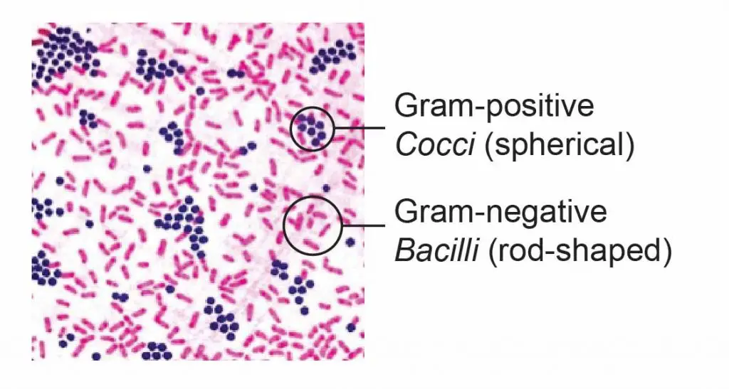
Module The Gram Stain Procedure click "start lab" From a liquid culture, take a loopful of bacteria emulsify it in a small drop of water or saline on the slide This should be a thin, not milky, suspension or it will not stain properly Air dry the slide This is done automatically in the virtual module To begin 1Escherichia coli Four different strains of Escherichia coli on Endo agar with biochemical slope Glucose fermentation with gas production, urea and H 2 S negative, lactose positive (with exception of strain D "late lactose fermenter";E coli S epidermidis Note Escherichia coli is a tiny pink (Gram) rod Staphylococcus epidermidis is a purple (Gram) sphere or coccus Draw a picture of a typical microscopic field and identify both Escherichia coli and Staphylococcus epidermidis Record this in the results section for this lab Colored pencils are available throughout the room
Description Precleaned glass slide with ground edges Professionally stained samples for best visualization Ideal for biology classrooms to explore structurefunction relationships as per NGSS standards Showing gram negative E Coli bacteria stained with carbolfuchsin Wholemount E Coli is aThe key difference between E coli and Pseudomonas aeruginosa is that E coli is a facultative anaerobic bacterial species that belongs to family Enterobacteriaceae and genus Escherichia, while P aeruginosa is an aerobic bacterial species that belongs to family Pseudomonadadaceae and genus Pseudomonas Both E coli and Pseudomonas aeruginosa are gramnegative, rodshaped and motile bacteriaFlood the sample with iodine and allow the slide to stand for about 1 minute


Gram Positive And Gram Negative Bacteria Structures Characterisitics


Mospace Umsystem Edu Xmlui Bitstream Handle 3 1 Sdm gst Pdf Sequence 12 Isallowed Y
The slide is stained with safranin for 1min, then rinse with water very gently until no color is coming from the water Blot dry with absorbent paper Place the slide on the microscope to view the sample The grampositive bacteria will stain dark blue or purpleOn Endo agar it looks like lactose negative)All four strains are mannitol positive (best seen in fig D), cellobiose negative (strains A, B)Description Precleaned glass slide with ground edges Professionally stained samples for best visualization Ideal for biology classrooms to explore structurefunction relationships as per NGSS standards Showing gram negative E Coli bacteria stained with carbolfuchsin Wholemount E Coli is a


What Does An E Coli Bacteria Look Like Under A Microscope Quora


Gram Stain Control Slides Scientific Device Laboratory
Heat fix the smear while holding the slide at one end, and by quickly passing the smear over the flame of Bunsen burner two to three times Procedure of Gram stain for E coli Cover the smear with crystal violet and allow it to stand for one minute Rinse the smear gently under tap waterStaphylococci (staphylococcus aureus)gram positive, spherical bacteris that have developed antibiotic resistant strains, 400x gram stain stock pictures, royaltyfree photos & images microscopic view of bacterial pneumonia bacterial pneumonia is a type of pneumonia caused by bacterial infection gram stain stock illustrations4 Gram Stain Protocol iii CSF and other body fluids requiring centrifugation Some laboratories may choose to use a Cytospin slide centrifuge to concentrate body fluids for s mear preparation This method has been recommended to increase Gram stain sensitivity and to decrease centrifugation and examination time for more rapid results


Observing Bacteria Under The Microscope Gram Stain Steps Rs Science


Jn Tm6z15kb1m
Many experiments in this course will utilize the Gram stain You should KNOW THE GRAM STAIN MATERIALS 1 Slant cultures of Escherichia coli, Micrococcus roseus, or other bacteria 2 Inoculating loops, Slides 3 Gram staining reagents in dropping bottles 4 Toothpicks 5 BHI plate from Experiment I PROCEDURE Heat Fixed Smear 1Spores – The Escherichia coli is a non–sporing bacterium Capsule – Capsules are present in some strains of the E coli which can easily be demonstrated using India ink preparation, appear as a clear halo in a dark background Gram Staining Reaction – Escherichia coli is a Gram ve (Negative) bacterium MORPHOLOGY OF ESCHERICHIA COLI (ECOLI)EXPERIMENT 1 A student is performing a Gram stain of a mixed culture of both E coli and S aureus and he forgets to decolorize with ethanol What should his slide look like?


Staphylococcus Aureus And Ecoli Under Microscope Microscopy Of Gram Positive Cocci And Gram Negative Bacilli Morphology And Microscopic Appearance Of Staphylococcus Aureus And E Coli S Aureus Gram Stain And Colony Morphology On Agar Clinical


Q Tbn And9gcswouuht13c4cxzkdgvwicdnmwhgkfjwlh40a Eerzzlpwjtmyt Usqp Cau
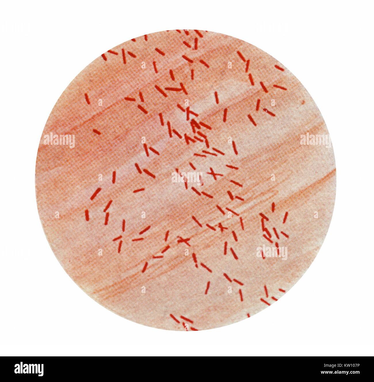
Assess Performance, Improve Accuracy QC1™ Gram Quality Control Slides allow your lab to objectively assess the performance of equipment, reagents, methods and techniques, while eliminating the need to maintain inhouse cultures of control organisms Slides are available in standard format (with only positive and negative controls) or with either one or three wells for patient specimens — making them ideal for the second and third shifts for supervisor review2 Morphology and Staining of Escherichia Coli E coli is Gramnegative straight rod, 13 µ x 0407 µ, arranged singly or in pairs (Fig 281) It is motile by peritrichous flagellae, though some strains are nonmotile Spores are not formed Capsules and fimbriae are found in some strains 3 Cultural Characteristics of Escherichia ColiGram Stain Tissue Trol Control Slides From Mouse Lung Tissue Gram Stain Of E Coli Bacterium A Gram Stain Of Shows Escherichia Coli Slide W M Science Lab Microbiology Supplies What Does An E Coli Bacteria Look Like Under A Microscope Quora Gram Stain Procedure In Microbiology


E Coli


Www Mccc Edu Hilkerd Documents Bio1lab3 Exp 4 000 Pdf
APPLICATION Newcomer Supply Gram Positive & Gram Negative Bacteria, Artificial Control Slides are for the positive histochemical staining of gram positive and gram negative bacteria in the same tissue section Escherichia coli and Staphylococcus aureus purchased from Remel Microbiology Products is used to produce the positive control tissue1 Escherichia coli a Fix a smear of Escherichia coli to the slide as follows 1First place a small piece of tape at one end of the slide and label it with the name of the bacterium you will be placing on that slide 2Using the dropper bottle of deionized water found in your staining rack, place 1/2 of a normal sized drop of water on a clean slide by touching the dropper to the slideNewcomer Supply Gram Positive & Gram Negative Bacteria, Artificial Control Slides are for the positive histochemical staining of gram positive and gram negative bacteria in the same tissue section Escherichia coli and Staphylococcus aureus purchased from Remel Microbiology Products is used to produce the positive control tissue


Gram Positive And Gram Negative Rods Microscopy Microbiology Microbiology Lab


Q Tbn And9gcqkye60ou Johpr02n Mbv1fferrjpdh Lnct7ymdf5qhyia1ld Usqp Cau
Part 5 Gram Stain Procedure Place slide with heat fixed smear on staining tray Gently flood smear with crystal violet and let stand for 1 minute Tilt the slide slightly and gently rinse with tap water or distilled water using a wash bottle Gently flood the smear with Gram's iodine and let stand for 1 minuteIt is rod shaped and is found in the human intestines The prepared microscope slide image of Bacillus Subtilis at left was captured at 400x magnification Learn more here E Coli E Coli under the microscope at 400x E Coli (Escherichia Coli) is a gramnegative, rodshaped bacteriumGramnegative Escherichia coli, the most common Gram stain qualitycontrol bacterium, is decolorized, and is only visible after the addition of the pink counterstain safranin (credit modification of work by Nina Parker)


What Does An E Coli Bacteria Look Like Under A Microscope Quora


Gram Stains Of Bacteria Ppt Video Online Download
Gram staining is a quick procedure used to look for the presence of bacteria in tissue samples and to characterise bacteria as Grampositive or Gramnegative, based on the chemical and physical properties of their cell walls The Gram stain should almost always be done as the first step in diagnosis of a bacteria infection The Gram stain is named after the Danish scientist Hans ChristianThe slide is stained with safranin for 1min, then rinse with water very gently until no color is coming from the water Blot dry with absorbent paper Place the slide on the microscope to view the sample The grampositive bacteria will stain dark blue or purpleE coli 1 PRESENTED BY NOMAN HAFEEZ KHOSA ABDUL SAMI FARHAN MISBAH SADIQ ANUM AJMAL Escherichia coli 2 2 Esherichia coli Gramnegative rod Facultative anaerobe Named for Theodor Escherich German physician (ca 15) Demonstrated that particular strains were responsible for infant diarrhea and gastroenteritis Normal flora of the mouth and intestine Protects the intestinal tract from bacterial


Photomicrographs


Microscope World Blog Microscopy Gram Staining
A slide is also not acceptable for examination if microorganisms that should be grampositive appear pink This may indicate that either the Gram's iodine was not applied or the slide was overdecolorized Staining grampositive and gramnegative control slides along with the patient's smear would confirm that proper staining technique was usedGram Staining Now that the slide has been heat fixed, you may now begin to stain the organism This is started by applying a few drops of crystal violet for 30 seconds, then rinsing with water for 5 seconds Next, cover the slide with Gram's Iodine and let sit for a full minute before rinsing with water for another 5 secondsThe prepared microscope slide image of Bacillus Subtilis at left was captured at 400x magnification Learn more here E Coli E Coli under the microscope at 400x E Coli (Escherichia Coli) is a gramnegative, rodshaped bacterium Most E Coli strains are harmless, but some serotypes can cause food poisoning in their hosts


Microbiologics Sl01 10 Gram Stain Control Slide


Gram Stain High Resolution Stock Photography And Images Alamy
The Gram negative organism is E coli These slides are intended to be used in conjunction with the GramPRO 1 and for manual staining Methyl Alcohol (Methanol) it is required that methanol fixation be used for clinical specimens or QC slides in preparation for the manual Gram stain or for slides run on QuickSlide™ automated Gram slideSingle, prepared microscope slide of Escherichia Coli (EColi) Wholemount, stained with Gram Stain EColi is a gramnegative bacterium, most commonly found in the digestive system of mammals While it is not typically harmful, the bacteria may become pathogenic Excellent for discussion about pathogenic vs nonpathogenic bacteria Slide measures 75mm (3") wide, 25mm (1") long and 006" in heightGram Stain Tissue Trol Control Slides From Mouse Lung Tissue Gram Stain Of E Coli Bacterium A Gram Stain Of Shows Escherichia Coli Slide W M Science Lab Microbiology Supplies What Does An E Coli Bacteria Look Like Under A Microscope Quora Gram Stain Procedure In Microbiology


5 Steps Of Gram Staining Procedure How To Interpret The Results New Health Advisor


Bacterial Characteristics Gram Staining Video Khan Academy
GramPRO 1 Gram stain QSlide™ PROBond slides are prepared from standard stock ATCC quality control organisms The Gram positive organism is Staphylococcus aureus The Gram negative organism is E coli These slides are intended to be used in conjunction with the GramPRO 1 and for manual staining* During heat fixing, avoid overheating the slide Gram Staining Place the slide on a staining rack and flood the smear with crystal violet for about one minute;We have cultures of E coli and Bacillus for you to gram stain This will give you gram and gram – controls to check your procedure against You can use 2 slides, 1 for each bacterium, or you can divide one slide in half and smear each bacterium on the divided slide There are also prepared, gram stained slides of bacteria of different shapes and sizes of bacteria to look at


Mechanical Penetration Of B Lactam Resistant Gram Negative Bacteria By Programmable Nanowires Science Advances


Gram Negative Bacteria Wikipedia
Description This slide of a smear of E coli is best viewed under a microscope with an oilimmersion objective three forms (bacillus, cocci and spirillum), smear, Gram stain Microscope slide AU$40 ex GST Bacillus subtilis, bacilli, smear, Gram stain Microscope slide AU$1410 ex GST Staphylococcus epidermidis, cocci, clusters, smear4 Aseptically transfer Escherichia coli (E coli) to circle on right Allow slide to heat fix S epi D Perform the Gram Stain 1 Take the heat fixed Gram slide to one of the staining racks located at the benches with sinks Flood each slide with crystal violet and allow to stand for one minute Rinse the slide with water 2As indicated in the student notes, the first slide that was prepared using aseptic technique and the Gram Staining method was a slide containing cultures of M luteus and E coli When examining this slide under the microscope at 1000X magnification, I observed some clusters and mostly doubles of coccishaped purple colonies
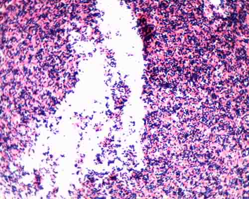


Gram Stain Microbiology Images Photographs


What Does Gram Positive Mean Fermup
Slightly tilt the slide and rinse with water (distilled or tap water) gently Avoid roughly washing the slide;Bio 112 Abstract Escherichia coli, Bacillus subtilis and Staphylococcus epidermidis were analyzed for this lab activity to determine their Gram Stain After the multilayered Gram Stain procedure each bacteria were classified as Grampositive or Gramnegative depending on their cell walls staining color The results showed that E coli stained pink and classified as GramnegativeFix a smear of either Escherichia coli, Pseudomonas aeruginosa, or Klebsiella aerogenes to the slide as follows 1 First place a small piece of tape at one end of the slide and label it with the name of the bacterium you will be placing on that slide


2
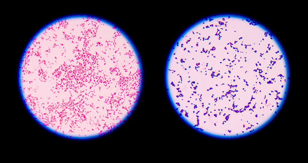


9 Gram Staining Best Practices Microbiologics Blog
E coli 1 Escherichia coli is a Gram negative, facultative anaerobic, rodshaped bacteria It is a commensal that is found inhabiting the lower intestine of warm blooded animals A small proportion of E coli strains are pathogenic The harmless strains produce vitamin K and prevent colonization of the intestine by pathogenic bacteria E coli is classified into serotypes based on cell wallEscherichia coli Four different strains of Escherichia coli on Endo agar with biochemical slope Glucose fermentation with gas production, urea and H 2 S negative, lactose positive (with exception of strain D "late lactose fermenter";SIMPLE STAIN and the GRAM STAIN In most microbiological staining procedures, the bacteria are first fixed to the slide by the heat fixed smear (Figure 1) In this procedure living, potentially pathogenic bacteria are smeared on the glass slide and allowed to air dry Then the slide is heated so that the bacteria are


Microscope World Blog Microscopy Gram Staining


Solved Lab Module 6 Gram Stain Lab Report Name Using T Chegg Com
E Coli Gram Stain And Cell Morphology E Coli Micrograph What Does An E Coli Bacteria Look Like Under A Microscope Quora Gram Stain Demonstration Slide 400x 2 A Slide Demonstrati Flickr S Epidermidis Gram Stain Escherichia Coli Slide W M Science Lab Microbiology SuppliesValidate proper staining of organisms when staining smears for gram positive organisms and improve work flow with clinical smears on the same slide with Gram Stain Control Slides wpb_animate_when_almost_visible { opacity 1;


Product Catalog


Photographic Print E Coli Bacteria Gram Negative Bacilli Gram Stain Lm X400 By Gladden Willis 24x18in Bacillus Photographic Print Microbiology


Academic Oup Com Labmed Article Pdf 32 7 368 Labmed32 0368 Pdf


A Novel Improved Gram Staining Method Based On The Capillary Tube


Q Slide Gram A Prepared Control Slide For The Qc Of The Gram Stain Procedure Has E Coli And S Aureus Pre Inoculated 5 Slides Per Box By Hardy Diagnostics Amazon Com Industrial Scientific
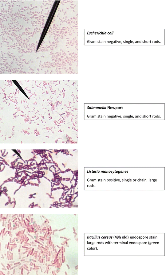


Staining Technology And Bright Field Microscope Use Springerlink


Bacteria Types Slide Separate Smears Gram Stain Science Lab Microbiology Supplies Amazon Com Industrial Scientific


Http Coltonanderson1 Weebly Com Uploads 2 4 3 0 Manual Pdf
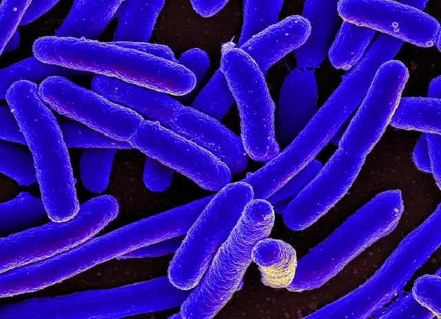


E Coli Under The Microscope Types Techniques Gram Stain Hanging Drop Method


Development Of A Standardized Gram Stain Procedure For Bacteria And Inflammatory Cells Using An Automated Staining Instrument Li Microbiologyopen Wiley Online Library


Mixed Culture 1 1 Of M Luteus Gram Positive Cocci And E Coli Download Scientific Diagram


Escherichia Coli Light Microscopy


Gram Staining Of Escherichia Coli Gram Staining Of Escherichia Coli Introduction Gram Staining Is Type Of Staining For Bacteria That Allows For An Individual To Studocu


Staining Microscopic Specimens Microbiology


A Control A Slide Showing Intact Rod Shaped E Coli 0157 H7 Cells And Download Scientific Diagram


Escherichia Coli Slide W M Science Lab Microbiology Supplies Amazon Com Industrial Scientific


We Need New Antibiotics For Gram Negative Not Gram Positive Bacteria Reflections On Infection Prevention And Control


Gram Stain Demonstration Slide 1 000x 2 A Slide Demonstra Flickr


Phenotypic Plasticity Of Escherichia Coli Upon Exposure To Physical Stress Induced By Zno Nanorods Scientific Reports


Gram Stain For Films Sigma Aldrich


Www Mccc Edu Hilkerd Documents Bio1lab3 Exp 4 000 Pdf


Gram Stains A Resource For Retrospective Analysis Of Bacterial Pathogens In Clinical Studies


Gram Stain Microbiology Images Photographs
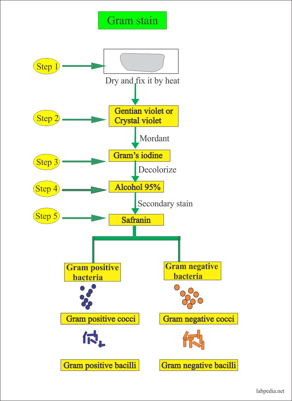


Gram Stain Gram Stain Procedure Labpedia Net


Gram Stain Staphylococcus Aureus E Coli Combined In Same Organ Control Histology Slides


Gram Staining Principle Procedure And Results Learn Microbiology Online


Http Learn Chm Msu Edu Vibl Content Gramstain Module Instructions Pdf
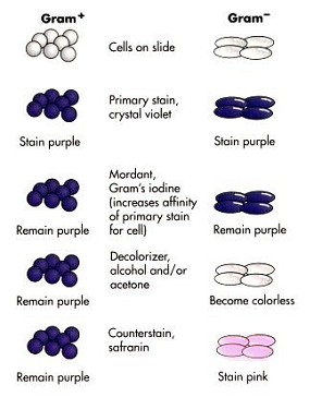


17 Gram Stain Biology Libretexts


Q Tbn And9gcqkye60ou Johpr02n Mbv1fferrjpdh Lnct7ymdf5qhyia1ld Usqp Cau


Gram Multi Tissue Artificial In Separate Tissues Quality Control Histology Slides
/gram-positive-staphylococcus-aureus-bacteria-541802136-57979cca5f9b58461f26eccc.jpg)


Gram Stain Procedure In Microbiology


E Coli Gram Stain Page 6 Line 17qq Com
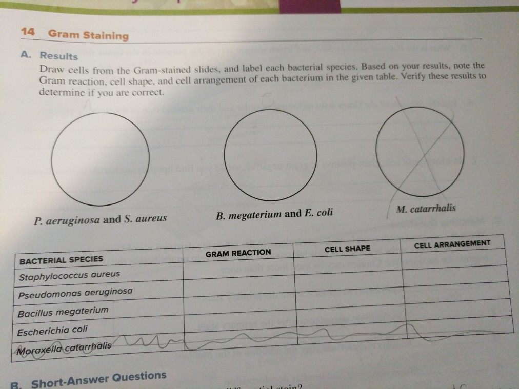


Solved 14 Gram Staining A Results Draw Cells From The Gr Chegg Com


Gram Stain Columbus County


Eiscoprepared Microscope Slide Escherichia Coli Smear Gram Stain Microbiology Fisher Scientific


Morphology Of Bacterial Cells


Asmscience Examination Of Gram Stains Of Urine


Product Catalog


Lab 3 Review Of Lab 2 Gram Staining Record Results On Pg 35 Gram Positive Purple Gram Negative Pink Bacillus Subtilis Escherichia Coli Klebsiella Ppt Download


Http Faculty Fiu Edu Gantarm Ex2gramstain Pdf


Gram Stain Introduction Principle Procedure Result And Interpretation


E Coli Gram Stain Page 1 Line 17qq Com


Solved Lab Module 6 Gram Stain Lab Report Name Using T Chegg Com


1b Demo


Gram Stain Wikipedia


Eiscoprepared Microscope Slide Escherichia Coli Smear Gram Stain Microbiology Fisher Scientific
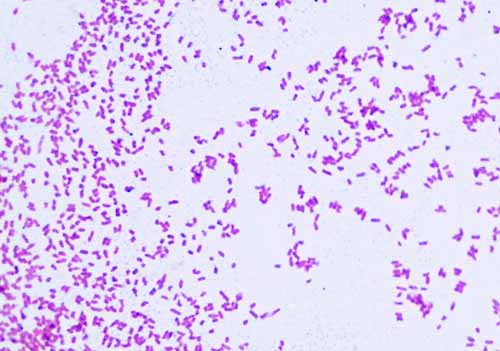


Gram Negative Bacteria Images Photos Of Escherichia Coli Salmonella Enterobacter


Microbiology Lab Exercise 10 Smear Preparation Exercise 11 Simple Staining Ex 12 Negative Staining Review Lab Results From Last Week Ex 8 Aseptic Technique Did You Successfully Get Bacteria Growing In Your Broth And Slant Ex 6 The


Gram Stain Demonstration Slide 400x 1 Marc Perkins Photography


Escherichia Coli Lecture


Eiscoprepared Microscope Slide Escherichia Coli Smear Gram Stain Microbiology Fisher Scientific
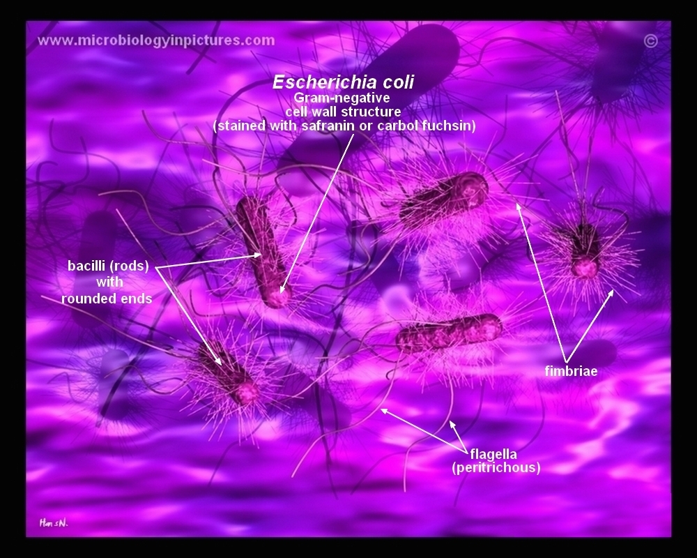


How E Coli Bacteria Look Like


Hardy Diagnostics Qs0700 Mckesson Medical Surgical
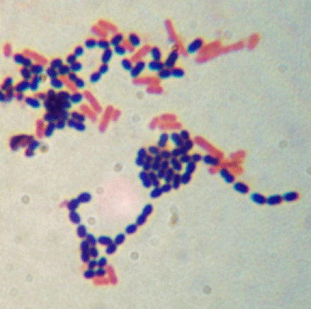


Welcome To Microbugz Gram Stain


Escherichia Coli Colony Morphology And Microscopic Appearance Basic Characteristic And Tests For Identification Of E Coli Bacteria Images Of Escherichia Coli Antibiotic Treatment Of E Coli Infections


Staining Microscopic Specimens Microbiology


Www Csus Edu Indiv R Rogersa Bio139 Holab9gramkoh Pdf
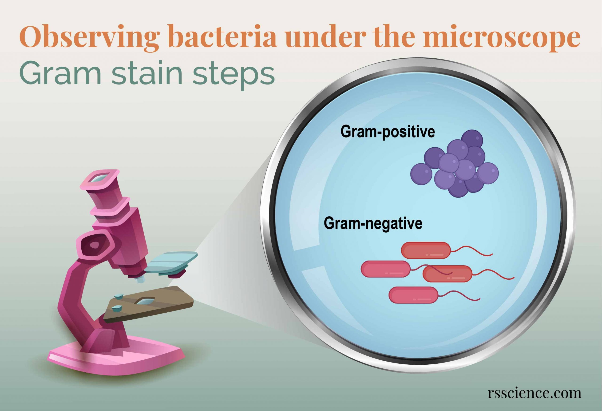


Observing Bacteria Under The Microscope Gram Stain Steps Rs Science


Gram Staining Slide Imagery Of Bacteria Cells In The Intestines Of Download Scientific Diagram


Gram Stain Tissue Trol Control Slides Mouse Lung Tissue Containing Staphylococcus Aureus And Escherichia Coli Sigma Aldrich


Pin On Microbiology


1


Lab 1


Bacteria Escherichia Coli Gram Stain Prepared Slide School Science Equipment


Gram Stain Demonstration Slide 400x 2 Marc Perkins Photography


What Does An E Coli Bacteria Look Like Under A Microscope Quora


Gram Stain Images Microbiology Stain Study Tools


Gram Stain Lab Report Record The Results Use The F Chegg Com


Laboratory Perspective Of Gram Staining And Its Significance In Investigations Of Infectious Diseases Thairu Y Nasir Ia Usman Y Sub Saharan Afr J Med
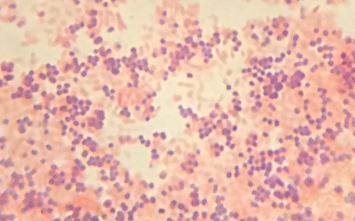


Staining Microscopic Specimens Microbiology
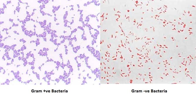


Gram Staining Principle Procedure Interpretation Examples And Animation
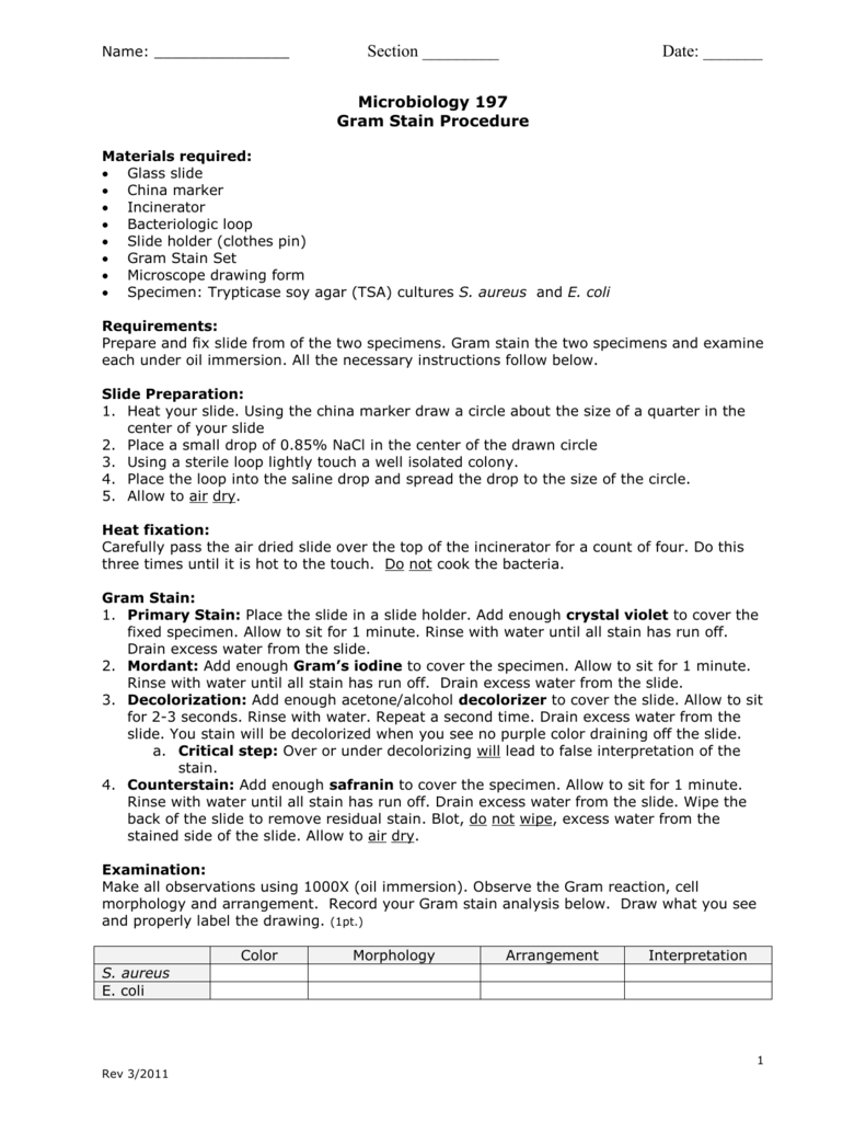


Gram Stain


E Coli Gram Stain Introduction Principle Procedure And Result Interpret


Asmscience Sputum Gram Negative Bacilli
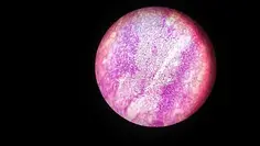


E Coli Under The Microscope Types Techniques Gram Stain Hanging Drop Method


Bacterial Slides Bacerial Stains Microbiology


コメント
コメントを投稿